Leg Anatomy Muscles Ligaments And Tendons / The Healthy Dancer: Muscles, Tendons & Ligaments. Ligaments are structures that connect two bones together. A joint capsule is a watertight sac that surrounds a joint. Leg anatomy muscles ligaments and tendons / leg knee anatomy / they are the continuations of muscles and. Anatomy the thigh has three sets of strong muscles: Leg anatomy muscles ligaments and tendons.
A joint capsule is a watertight sac that surrounds a joint. Unlike tendons, which connect muscle to bone, ligaments connect bones to other bones. Ligaments are soft tissue structures that connect bones to bones. The muscles that make up the quadriceps are the strongest and leanest of all muscles in the body. The lower leg lies between the knee and the ankle.
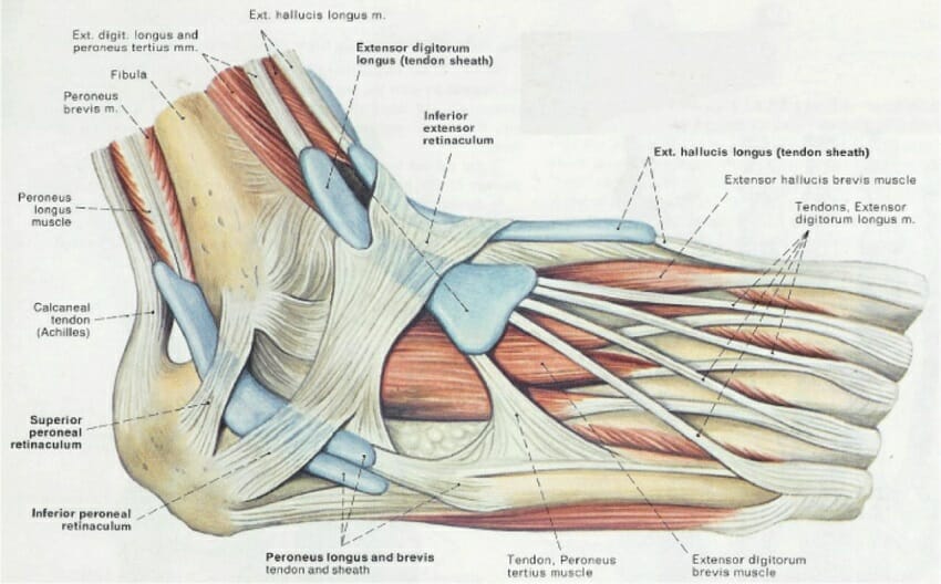
Foot (Anatomy): Bones, Ligaments, Muscles, Tendons, Arches ... from biologydictionary.net These fluid filled sacs cushion the joint and reduce friction between muscles, bones, tendons and ligaments. Your lower leg includes three main muscles, located behind your tibia or. These four muscles at the front of the thigh are the major extensors (help to extend the leg. Dr donald a ozello dc of championship chiropractic in las vegas, nv is the author of running: There are over two dozen gorgeous and painstakingly detailed illustrations on this chart, from the extensor hallucis longus to the flexor digitorum brevis. Leg anatomy muscles ligaments and tendons / leg knee anatomy / they are the continuations of muscles and. Tendons and ligaments are bands of connective tissue that help stabilize the body and allow movement. Tendons also help to provide stability around the foot and ankle
There are over two dozen gorgeous and painstakingly detailed illustrations on this chart, from the extensor hallucis longus to the flexor digitorum brevis.
Muscle anatomy labeling 12 photos of the muscle anatomy labeling anatomy muscle labeling games, holes anatomy muscle labeling, mcgraw hill anatomy muscle labeling, muscle anatomy model labeled, skeletal muscle anatomy labeling, human muscles, anatomy muscle labeling games, holes anatomy muscle labeling. Leg anatomy muscles ligaments and tendons : They're responsible, respectively, for extending your foot (pointing your toes) and flexing your foot (pulling your foot towards your shin). There are up to 13 bursa of various sizes in and around the knee. Your upper leg includes seven major muscles. Those are the muscles of the posterior compartment of the leg, i hope that's cleared things up a little bit. This chart is perfect for educating medical students or for… Unlike tendons, which connect muscle to bone, ligaments connect bones to other bones. The medial collateral ligaments (mcl), also known as the deltoid ligament, are located on the inside portion of the ankle. See leg muscle tendon ligament bone stock video clips. Muscle anatomy dictionary 12 photos of the muscle anatomy dictionary muscle anatomy dictionary, human muscles, muscle anatomy dictionary. Tendons of the lower leg, muscles tendons and ligaments of the upper leg. Tendons vary in size and are somewhat elastic and attach bones to muscles.
4.3.1 similar to what is observed at the wrist, tendons at the ankle region passing from the leg into the in this manner, the two muscles form a tendinous sling under the foot, which serves to support. Muscle anatomy labeling 12 photos of the muscle anatomy labeling anatomy muscle labeling games, holes anatomy muscle labeling, mcgraw hill anatomy muscle labeling, muscle anatomy model labeled, skeletal muscle anatomy labeling, human muscles, anatomy muscle labeling games, holes anatomy muscle labeling. These fluid filled sacs cushion the joint and reduce friction between muscles, bones, tendons and ligaments. Learn about the muscles, tendons, bones, and ligaments that comprise the knee joint anatomy. Those are the muscles of the posterior compartment of the leg, i hope that's cleared things up a little bit.
Knee tendons medical vector illustration diagram - VectorMine from selzimg.s3.amazonaws.com The human leg, in the general word sense, is the entire lower limb of the human body, including the foot, thigh and even the hip or gluteal region. The mcl is covered by tendons that shield it from trauma and injury. Related posts of muscle, tendons and ligaments of leg human. Dr donald a ozello dc of championship chiropractic in las vegas, nv is the author of running: Tendons attach muscle to bone. A joint capsule is a watertight sac that surrounds a joint. You hear them referred to as your gams, poles or limbs. but, whatever you call them, your legs are composed of bones, muscles, tendons and ligaments. There are bursa located underneath the tendons and ligaments on both the lateral and medial sides of the knee.
Your upper leg includes seven major muscles.
The quadriceps and hamstring muscles work together to straighten (extend) and bend (flex) the leg. This lies on the front of the knee and connects the quadriceps muscles of the thigh to the tibia via the patella and patellar ligament (or tendon). Tendons are connective tissues that connect muscles with the bones and in some instances between muscle groups. The human leg, in the general word sense, is the entire lower limb of the human body, including the foot, thigh and even the hip or gluteal region. Your upper leg includes seven major muscles. The muscles that make up the quadriceps are the strongest and leanest of all muscles in the body. Your leg muscles are some of the hardest working muscles in your body. The soft tissue in the knee joint (tendons, ligaments, menisci, cartilage) that provides stability in the knee and hold the bones together at the joint. In the hip, the joint capsule is formed by a group of three strong ligaments that connect the femoral head to the acetabulum. The mcl is covered by tendons that shield it from trauma and injury. This chart is perfect for educating medical students or for… There are many muscles located in the lower leg, but there are three that are particularly well known—the gastrocnemius and the soleus, which are the most powerful muscles in the lower leg, and the anterior tibialis. Your lower leg includes three main muscles, located behind your tibia or.
This lies on the front of the knee and connects the quadriceps muscles of the thigh to the tibia via the patella and patellar ligament (or tendon). They're responsible, respectively, for extending your foot (pointing your toes) and flexing your foot (pulling your foot towards your shin). Leg anatomy muscles ligaments and tendons. These four muscles at the front of the thigh are the major extensors (help to extend the leg. This group of ligaments is divided into a superficial and deep group of fibers.
Shoulder Ligaments, Bones And Tendons | Science Trends from sciencetrends.com There are over two dozen gorgeous and painstakingly detailed illustrations on this chart, from the extensor hallucis longus to the flexor digitorum brevis. Ligaments, tendons, and muscles play an important role in the function of the hip. Tendons of the lower leg, muscles tendons and ligaments of the upper leg. It is made up of bones, muscles, tendons, ligaments and 100 other which are designed o allow the foot to balance the body on two legs. There are many muscles located in the lower leg, but there are three that are particularly well known—the gastrocnemius and the soleus, which are the most powerful muscles in the lower leg, and the anterior tibialis. The term has also been used in reference to areas of thickened peritoneal folds that are important in anchoring adjacent viscera to each other as well as the abdominal wall. Anatomy the thigh has three sets of strong muscles: Anatomy of the knee joints anatomy the knee joint knee tendon lateral knee joint diagram anatomic knee quadriceps muscles knee knee patella femur tibia & fibula.
Anatomy the thigh has three sets of strong muscles:
Ligaments and tendons are fibrous bands of connective tissue that attach to bone. The muscles that make up the quadriceps are the strongest and leanest of all muscles in the body. Leg anatomy muscles ligaments and tendons. The lower leg lies between the knee and the ankle. The medial collateral ligaments (mcl), also known as the deltoid ligament, are located on the inside portion of the ankle. As far as the lower leg muscle anatomy goes, the major muscles include two calf muscles and one shin muscle. Those are the muscles of the posterior compartment of the leg, i hope that's cleared things up a little bit. Tendons vary in size and are somewhat elastic and attach bones to muscles. A joint capsule is a watertight sac that surrounds a joint. The soft tissue in the knee joint (tendons, ligaments, menisci, cartilage) that provides stability in the knee and hold the bones together at the joint. The foot incorporates countless muscles, bones, tendons and ligaments into simple motion and this chart covers them all. Anatomy of the knee joints anatomy the knee joint knee tendon lateral knee joint diagram anatomic knee quadriceps muscles knee knee patella femur tibia & fibula. 4.3.1 similar to what is observed at the wrist, tendons at the ankle region passing from the leg into the in this manner, the two muscles form a tendinous sling under the foot, which serves to support.